Imaging Technologies
The objective of the CERIMED project is to bring a significant breakthrough in sensitivity, spatial resolution and multimodality capability of presently available imaging devices. The impressive technological developments in several areas of applied physics, in particular to answer the challenge of a new generation of particle physics detectors and of the development of an information based society open the way to a major breakthrough in the performance of presently available imaging tools. The ultimate goal is to develop a high resolution, high sensitivity multi-energy X-ray/gamma detection module compatible with very high imaging magnetic field. Such a detector head will provide a quantum step towards truly multimodality for in vivo molecular imaging involving MRI, CT, PET and/or SPECT able to co-register the anatomy with the metabolic function quantitatively. CT is important in this regards, since it can be used to scale up the attenuation map and obtain absolute activity measurement in the case of PET.
Impressive progress have been made in the recent years in conversion materials for the detection of X-rays or Gamma-rays.
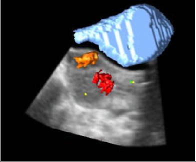
A combined ultrasound-MRI image
Typically, the detector requirements for PET or SPECT are high detection efficiency for sensitivity, fast timing for random event rejection and good energy resolution for scatter rejection. The optimisation of all of these parameters has led to the use of scintillation-based detectors, in which the energy of the incident X-ray or Gamma-ray is converted into a pulse of visible light. Some recent improvements have been made with the substitution of new brighter and faster crystals such as LSO and GSO (Lutetium and Gadolinium Silicates) for the traditional BGO (Bismuth Germanate) scintillator, whose relatively low light output and slow decay time put limits on the performance, despite its high density. A great deal of active research is on going to discover and produce at an industrial scale new scintillating materials like LuAP (Lutetium Aluminium Perovskite) that provide improved all-round performance. The crystal is coupled to a photo-detector, in which the scintillation light is converted into an electrical signal. This photo-detector is either a vacuum tube of the photomultiplier type, in which the scintillation light from the crystal extracts electrons from a photocathode, which are then accelerated in a strong electrostatic field and amplified on a series of dynodes before being collected on the anode of the tube, or a solid state diode or avalanche photo-diode, where electron-hole pairs are produced in the material by the incident light and migrate (with or without amplification) in the presence of an electrical field to electrodes deposited on two opposite faces of the detector. Finely segmented and efficient photo-detectors are mandatory for converting into electronic pulses the light signals produced in crystals of smaller and smaller sizes. Much progress has been made recently in the production of high gain and very compact multichannel photo-detectors of both types.
Another approach is the direct detection of X-rays or Gamma-ray radiation through the production of electron-hole pairs in the material in a similar way as for photodiodes. The problem here is related to the collection efficiency of electrons in materials which need to be dense and thick enough to convert the high energy incident radiation with a sufficient probability.
However impressive progress are going on in the production of new semiconductor material like CdZnTe or HgI2, which appear as very promising candidate for such an application to PET or SPECT cameras.
The direct integration on each pixel of the front end electronics (low noise preamplifier, shaper, analog logic) opens the door to more compact and cheaper detector heads. The compactness is here an essential feature for the integration of a truly multimodal device. The fact that such photo-detectors are generally immune to even strong magnetic fields is of course of great importance for MRI compatibility. Scintillators or direct conversion materials both require the design of complex and delicate mixed-signal low-noise amplifier and signal processing circuits to readout photoconductor and photodetector signals with the minimum possible noise at the required speed. These requirements are driven by the small signal available from the detector and by the fast signal processing needed to minimize the coincidence time window in the case of PET, or to maximize the counting frequency for CT taking into account the signal characteristics of photodetector and photoconductor. An important limitation of modern medical imaging equipment is on the data acquisition logic. The very high challenge of enormous data taking rates in High Energy Physics experiments has led several particle physics laboratories all over Europe to develop modern integrated technologies including pipelining and multi-level trigger logic. The front-end electronics chip should therefore integrate sophisticated signal processing and parallel, pipelined architectures to handle the enormous data stream and generate trigger primitives for the camera detection head. New progress in highly integrated electronics with very high bandwidth will lead to compact detector modules with a high pixellization, which will be better suited to the multimodality requirement.
Monte Carlo simulation is an essential tool in medical imaging to assist in the design of new medical imaging devices, assess new implementations of image reconstruction algorithms and/or scatter correction techniques, and optimise scan protocols. GATE, the GEANT4 Application for Tomographic Emission, encapsulates the GEANT4 libraries developed by the particle physics community in order to achieve a modular, versatile, scripted simulation toolkit adapted to the field of nuclear medicine. In particular, GATE allows the users to describe time-dependent phenomena such as detector movements or source decay kinetics, thus allowing to simulate time curves under realistic acquisition conditions. GATE is developed within the OpenGATE Collaboration with the objective to provide the academic community with a free software, general-purpose, GEANT4-based simulation platform for emission tomography.
This collaboration of software experts is also at the cutting edge of modern image reconstruction techniques, an essential tool for a total and easy exploitation of the complex information contained in multimodal systems.
___________________________________________________________________________________