ClearPET/XPAD
The ClearPET/XPAD project, development of small animal PET/CT scanner Centre de Physique des Particules de Marseille (CPPM), CNRS-IN2P3 and Université de la Méditerranée.
In contrast to X-ray computerized tomography (CT) that allows to imaging the mass density of living tissues, positron emission tomography (PET) is imaging gamma rays directly emitted from the tissues. In this case, the emission of gamma rays results from the decay of radioactive nuclei used to label a radiopharmaceutical injected to the patient. Thus, in the contrary to transmission tomography using an external X-ray tube, emission tomography images the function rather than the structure of living tissues.
The development of combined PET/CT imaging systems led to a rapid expansion of this technique in clinical routine. Similarly, the development of dedicated PET scanners and micro-CT scanners for small animals pleads for joining these two modalities in a common gantry. However, like with clinical PET/CT, the juxtaposition of both modalities does not allow for extrapolating the exact position of the animal during the PET scan. Therefore, we intend to combine the ClearPET detectors [1] with the XPAD3 X-ray hybrid pixel detector [2,3] in a common rotating gantry. To achieve this goal, we redesigned the partial ring arrangement of the ClearPET detectors. As a result, both detection systems will be merged together with an X-ray tube in a fully integrated PET/CT device that will make it possible to acquire simultaneous emission and transmission scans for mice.
The small animal ClearPET scanners have been developed during the past years within the Crystal Clear Collaboration [4]. All scanners use the same detector technology based on the combination of crystal matrices coupled with multi- anode photomultiplier tubes (MaPMTs). Since the sensitive area of the MaPMTs is smaller than their outer dimension, there is always an empty space between two axially adjacent detector blocks. This leads to empty slices in the sinograms, which cause loss of scanner sensitivity and severe artefacts in the images. In its present configuration, the ClearPET demonstrator consists of 18 LSO/LuYAP phoswich detector heads [1] arranged in three adjacent partial rings. Each ring consists of 6 detector modules mounted asymmetrically so that only two modules are facing each other [5].
A new detector arrangement that does not have axial gaps between adjacent rings is achieved by avoiding axially adjacent detector modules. Then, thanks to the rotation of the detectors around the FOV, the complete set of 3D sinograms can be addressed completely. Sensitivities are evaluated for a 15 cm length and 12 cm diameter cylindrical phantom using the GATE Monte Carlo simulation platform [6].
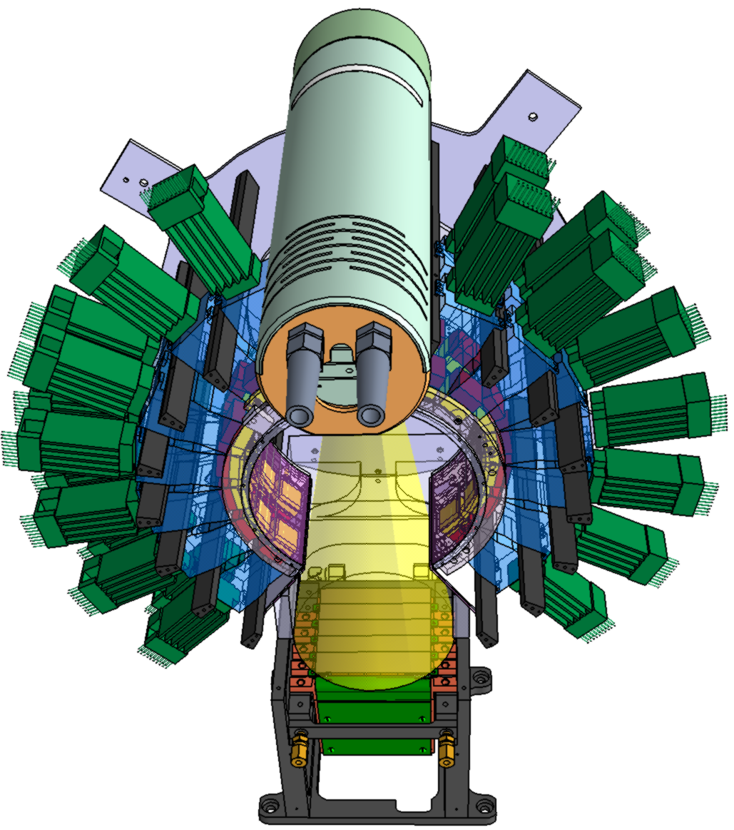
The layout of the ClearPET/XPAD dual-modality scanner mounted on a rotating gantry is shown in Figure 1. A detector configuration is implemented with three partial rings of 7 detector modules. It provides a PET modality axial FOV of 54 mm and a transverse FOV of 110 mm. The CT components (an X-ray tube from RTW with 50 μm focal spot size and the 118x76 mm2 XPAD3 X-ray imager with 130x130 μm2 pixels) are positioned within the PET detector ring. The microCTmodality has an axial FOV of 59 mm and a transverse FOV of 38 mm with a magnification by a factor 2. The X-ray cone beam passes through the shared FOV without irradiating the PET detectors directly.
Hence, the ClearPET/XPAD system will allow for simultaneous scanning of small animals. Impact of scattered X-ray on the LSO/LuYAP phoswich detectors modules are currently being studied using GATE and experimental measurements. This work is supported by the ANR Programme « Chaire d'excellence 2005 » under grant No. ANR-05-CEXC-01 and by the Conseil Régional PACA. It was labelled in 2006 by the pole of competitiveness 3Photonique/ systèmes complexes d’optique et d’imagerie”, in collaboration with the IBDML, the APHM, Créatis, Vegatec sarl and raytest France sarl.

Figure: The CLEARPET/XPAD design
REFERENCES
[1] J.-B. Mosset et al. Development of an optimized LSO/LuYAP phoswich detector head for the Lausanne ClearPET demonstrator. IEEE Trans Nucl Sci 53 (2006) 25-29.
[2] P. Delpierre et al. XPAD: A photons counting pixel detector for material science and small-animal imaging. Nucl Instr Meth A 572 (2007) 250-new. [3] P. Pangaud et al. XPAD3: A NEW photon counting chip for Xray CT-scanner. Nucl Instr Meth A 571 (2007) 321-324.
[4] K. Ziemons et al. The ClearPETTM project: development of a 2nd generation high-performance small animal PET scanner. Nucl Instr Meth A 537 (2005) 307-311.
[5] M. Rey et al. Count rate performance study of the Lausanne ClearPET scanner demonstrator. Nucl Instr Meth A 571 (2007) 207-210. [6] S. Jan et al. GATE : a simulation toolkit for PET and SPECT. Phys Med Biol 49 (2004) 4543-4561.
___________________________________________________________________________________
