The role of Imaging in modern medicine
New therapeutic strategies are entering the world of major diseases. These impose to acquire as fast as possible all the information on the pathological status of the patient in order to start adapted therapeutics and therefore to minimize the handicap. This is true for neurological and psychiatric diseases but also for the treatment of inflammatory diseases, such as rheumatologic inflammation, and for cancer.
Moreover, the non-invasive determination of the molecular signature of cancers in the early stage of their development, or even before the tumour growth, will help to target the therapeutic strategy and to reduce considerably the number of unnecessary biopsies.
For many years, physicians relied on the use of anatomical imaging to non-invasively detect tumours and follow up their growth. Functional imaging such as bone or thyroid scintigraphy and more recently positron emission tomography (PET) using 18FDG for example, has provided more information for tumour staging. With the development of new imaging probes and "smart probes", imaging provides cellular protein and signal-pathway identification, and represents a new field called "molecular imaging". A number of Centres have established collaborations with chemistry laboratories and are producing an increasing amount of molecular probes dedicated to imaging but also to tumour therapy. The molecular phenotype of cells composing the tumour can lead to tailored therapies. This tumour phenotype can be determined ex-vivo on tissue samples. Molecular imaging should allow performance of an in-vivo tumour phenotyping by a rationalized use of specific imaging probes. This molecular profiling could already be envisioned in the very near future for some specific tumours overexpressing peptide hormone receptors such as breast and prostate cancers, and should become widely developed.
Therefore, it represents a major breakthrough to provide the medical community with an integrated "one-stop-shop" molecular profiling imaging device which could detect tracers dedicated to Single Photon Emission Computed Tomography (SPECT) or PET, as well as Magnetic Resonance Imaging (MRI), or Xray-Computed Tomography (CT) contrast agents.
Furthermore, since functional imaging allows the assessment of biochemical pathways, it will also provide accurate tools for experimental research. As an example, the genomic alphabet has been decoded but its dynamic expression, its grammar, remains to be studied and understood. In-vivo molecular imaging of gene expression is now within reach through the development of more and more elaborated molecular probes as well as of sophisticated techniques to significantly improve the performances of modern imaging devices.
Drug development can also take advantage of technical progress in imaging technologies, like quantitative positron emission tomography in small animals, to determine drug pharmacokinetics and whole body targeting to tissues of interest. Moreover, the combination of functional imaging with a high resolution anatomical method such as MRI (Magnetic Resonance Imaging) and/or X-Ray CT (Computed Tomography) will considerably enhance the possibility of determining the long term efficiency of a drug on basic pathological processes such as inflammation, blood flow, etc. In particular the expected progress on sensitivity, timing and spatial resolution, coupled with a true multi-modality registration, will allow to explore the activity of a drug candidate or other essential patho-physiological processes of disease models in animals, like for instance cancer or adverse inflammatory effects.
The new trends in modern imaging is to develop adequate detector modules and radiotracers which can register simultaneously anatomical and functional information, and therefore fruitfully determine the molecular profiling.
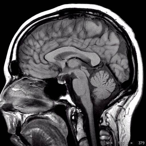
MRI image of a human skull and brain
___________________________________________________________________________________